MOLEQULE-ON reagents and kits are essential tool in research and various field of science, which enable accurate experiments and tests. Our products are integrated with newest technologies to meet the demand of a wide range of laboratories. The list includes PCR and qPCR reagents, electrophoresis, enzymes, nucleic acid extraction and purification kits, colorimetric and ELISA kits and buffers.
Western Blot
MOLEQULE-ON is a provider of Biochemistry, Microbiology and Biotechnology products. It Includes equipment, kits, PCR reagents, dehydrated culture media and plasticware. Our company is focused on customer satisfaction striving to gain the reputation of a reliable supplier of user-friendly laboratory tools.
Protein Staining Solution
Ponceau S Protein Staining Solution
Western chemiluminescence Substrate
PBS Buffer
Easy Protein Gels
MQ Western One Step Blocker
MQ Western Stripping Buffer
Tris Buffered Saline Buffer, TBS, (20X)
MQ Nano - Tech Protein Staining Solution
Description
MQ
Nano
-
Tech
Protein
Staining
Solution,
improved
by
nano
-
technology,
is
a
ready
-
to
-
use
protein
staining
solution
for
SDS - PAGE
gels.
Its
next
generation
formula
offers
a
faster
protein
detection,
higher
sensitivity
and
there
is
no
need
for
destaining.
Also,
the
washing
step
can
be
omitted.
In
the
absence
of
hazardous
substances
such
as
methanol
and
acetic
acid,
it
is
considered
to
be
safe
and
environmentally
friendly.
MQ
Nano
-
Tech
Protein
Staining
Solution
is
also
compatible
with
mass
spectrophotometry.
Features
1. Car
background
2. Fast
staining
3. No
need
for
destaining
4. No
washing
step
needed
5. No
overstaining
issue
6. No
hazardous
materials
inside
7. No
need
to
microwave
or
heat
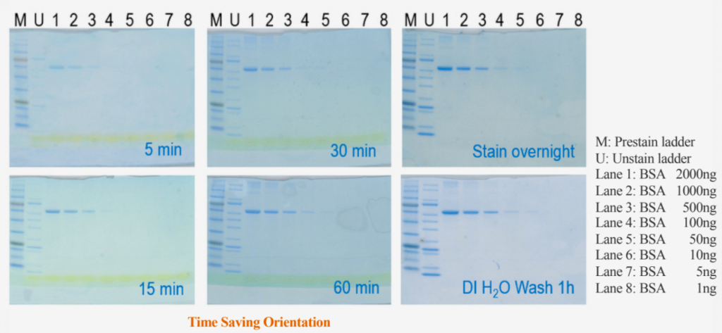
Prestained
protein
ladder,
unstained
protein
ladder,
and
BSA
were
prepared
and
applied
in
electrophoresis.
After
running
SDS
-
PAGE
(4 - 20%),
gel
was
removed
from
the
cassette
then
proceed
to
submerge
the
gel
in
proper
amount
of
MQ
Nano
-
Tech
Protein
Staining
Solution,
enough
to
cover
the
gel.
The
staining
box
was
lightly
agitated
for
5
minutes
to
over
night
at
room
temperature.
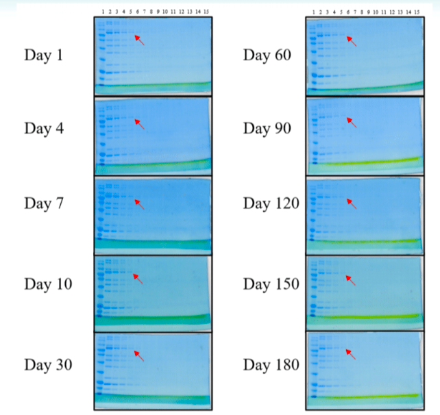
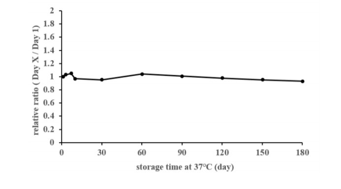
Analysis
of
85
kDa
band
of
lane
5
(red
arrow)
was
done,
The
band
intensity
between
different
storage
time
at
37°C
was
compared.
The
variation
is
under
10%.
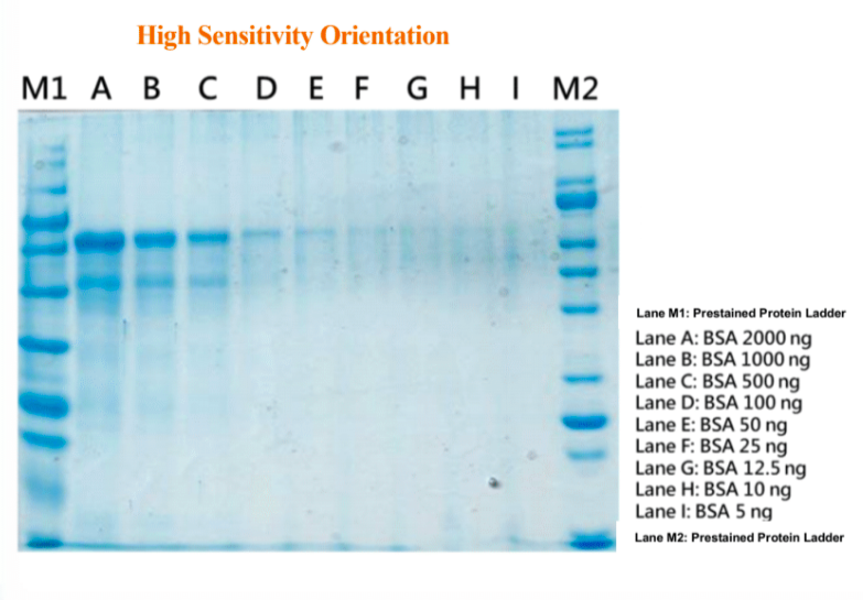
MQ
Nano
-
Tech
Protein
Staining
Solution
demonstrates
the
high
sensitivity
detection
could
be
down
to
10
ng
(Lane
H).
Prestained
protein
ladder,
unstained
protein
ladder,
and
serial
diluted
BSA
were
prepared
and
applied
in
electrophoresis.
After
running
10%
homemade
gel
(0.75 mm
thickness),
please
remove
the
gel
from
the
cassette
then
proceed
to
submerge
the
gel
in
a
proper
amount
of
MQ
Nano
-
Tech
Protein
Staining
Solution,
enough
to
cover
the
gel.
Lightly
agitate
the
staining
container
at
room
temperature
when
staining
for
30
hrs.
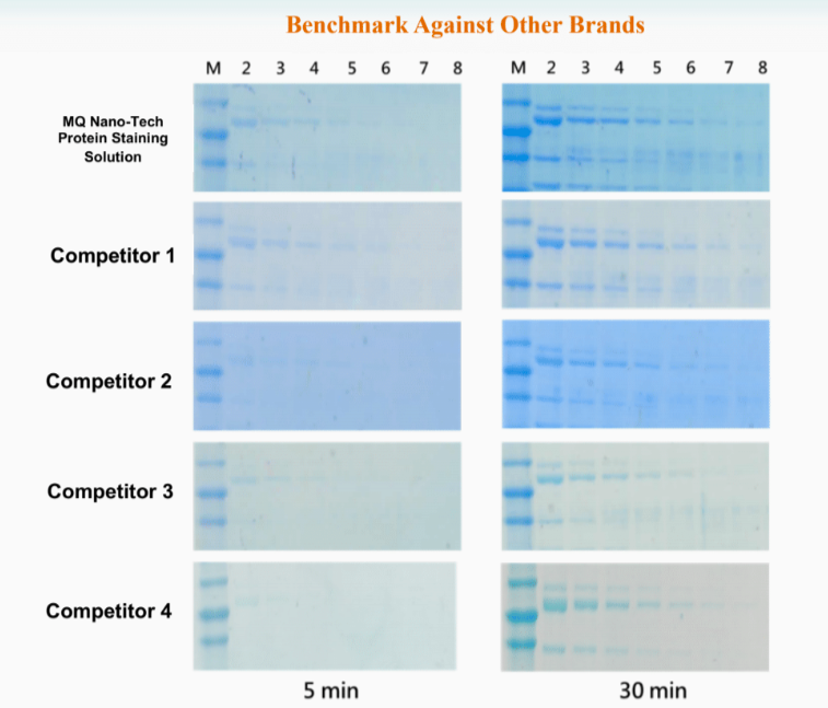
MQ
Nano
-
Tech
Protein
Staining
Solution
presents
the
equivalent
or
even
superior
performance
on
fast
signals
and
clear
background
compared
with
other
brands.
Lightly
agitate
the
staining
box
for
5
to
30
minutes
at
room
temperature.
MQ
Nano
-
Tech
Protein
Staining
Solution,
improved
by
nano
-
technology,
is
available
as
5X
concentrate
used
for
SDS-PAGE
gels.
Its
next
generation
formula
offers
a
faster
protein
detection,
higher
sensitivity
and
there
is
no
need
for
destaining.
Also,
the
washing
step
can
be
omitted.
In
the
absence
of
hazardous
substances
such
as
methanol
and
acetic
acid,
it
is
considered
to
be
safe
and
environmentally
friendly.
MQ
Nano-Tech
Protein
Staining
Solution
is
also
compatible
with
mass
spectrophotometry.
Features
1. Clear
background
2. Fast
staining
3. No
need
for
destaining
4. No
washing
step
needed
5. No
overstaining
issue
6. No
hazardous
materials
inside
7. No
need
to
microwave
or
heat
Prestained
protein
ladder,
unstained
protein
ladder,
and
BSA
were
prepared
and
applied
in
electrophoresis.
After running
SDS -PAGE(4-20%),
gel
was
removed
from
the
cassette
then
proceed
to
submerge
the
gel
in
proper
amount
of
MQ
Nano - Tech
Protein
Staining
Solution,
enough
to
cover
the
gel.
The
staining
box
was
lightly
agitated
for
5
minutes
to
over
night
at
room
temperature.
Analysis
of
85
kDa
band
of
lane
5
(red
arrow)
was
done,
The
band
intensity
between
different
storage
time
at
37°C
was
compared.
The
variation
is
under
10%.
MQ
Nano
-
Tech
Protein
Staining
Solution
demonstrates
the
high
sensitivity
detection
could
be
down
to
10
ng
(Lane
H).
Prestained
protein
ladder,
unstained
protein
ladder,
and
serial
diluted
BSA
were
prepared
and
applied
in
electrophoresis.
After
running
10%
homemade
gel
(0.75
mm
thickness),
please
remove
the
gel
from
the
cassette
then
proceed
to
submerge
the
gel
in
a
proper
amount
of
MQ
Nano
-
Tech
Protein
Staining
Solution,
enough
to
cover
the
gel.
Lightly
agitate
the
staining
container
at
room
temperature
when
staining
for
30
hrs.
MQ
Nano
-
Tech
Protein
Staining
Solution
presents
the
equivalent
or
even
superior
performance
on
fast
signals
and
clear
background
compared
with
other
brands.
Lightly
agitate
the
staining
box
for
5
to
30
minutes
at
room
temperature.
MQ
Nano
-
Tech
Protein
Staining
Solution,
improved
by
nano
-
technology,
is
a
ready
-
to
-
use
protein
staining
solution
for
SDS - PAGE
gels.
Its
next
generation
formula
offers
a
faster
protein
detection,
higher
sensitivity
and
there
is
no
need
for
destaining.
Also,
the
washing
step
can
be
omitted.
In
the
absence
of
hazardous
substances
such
as
methanol
and
acetic
acid,
it
is
considered
to
be
safe
and
environmentally
friendly.
MQ
Nano
-
Tech
Protein
Staining
Solution
is
also
compatible
with
mass
spectrophotometry.
Features
1. Car
background
2. Fast
staining
3. No
need
for
destaining
4. No
washing
step
needed
5. No
overstaining
issue
6. No
hazardous
materials
inside
7. No
need
to
microwave
or
heat
Prestained
protein
ladder,
unstained
protein
ladder,
and
BSA
were
prepared
and
applied
in
electrophoresis.
After
running
SDS
-
PAGE
(4 - 20%),
gel
was
removed
from
the
cassette
then
proceed
to
submerge
the
gel
in
proper
amount
of
MQ
Nano
-
Tech
Protein
Staining
Solution,
enough
to
cover
the
gel.
The
staining
box
was
lightly
agitated
for
5
minutes
to
over
night
at
room
temperature.
Analysis
of
85
kDa
band
of
lane
5
(red
arrow)
was
done,
The
band
intensity
between
different
storage
time
at
37°C
was
compared.
The
variation
is
under
10%.
MQ
Nano
-
Tech
Protein
Staining
Solution
demonstrates
the
high
sensitivity
detection
could
be
down
to
10
ng
(Lane
H).
Prestained
protein
ladder,
unstained
protein
ladder,
and
serial
diluted
BSA
were
prepared
and
applied
in
electrophoresis.
After
running
10%
homemade
gel
(0.75 mm
thickness),
please
remove
the
gel
from
the
cassette
then
proceed
to
submerge
the
gel
in
a
proper
amount
of
MQ
Nano
-
Tech
Protein
Staining
Solution,
enough
to
cover
the
gel.
Lightly
agitate
the
staining
container
at
room
temperature
when
staining
for
30
hrs.
MQ
Nano
-
Tech
Protein
Staining
Solution
presents
the
equivalent
or
even
superior
performance
on
fast
signals
and
clear
background
compared
with
other
brands.
Lightly
agitate
the
staining
box
for
5
to
30
minutes
at
room
temperature.
MQ
Nano
-
Tech
Protein
Staining
Solution,
improved
by
nano
-
technology,
is
available
as
5X
concentrate
used
for
SDS-PAGE
gels.
Its
next
generation
formula
offers
a
faster
protein
detection,
higher
sensitivity
and
there
is
no
need
for
destaining.
Also,
the
washing
step
can
be
omitted.
In
the
absence
of
hazardous
substances
such
as
methanol
and
acetic
acid,
it
is
considered
to
be
safe
and
environmentally
friendly.
MQ
Nano-Tech
Protein
Staining
Solution
is
also
compatible
with
mass
spectrophotometry.
Features
1. Clear
background
2. Fast
staining
3. No
need
for
destaining
4. No
washing
step
needed
5. No
overstaining
issue
6. No
hazardous
materials
inside
7. No
need
to
microwave
or
heat
Prestained
protein
ladder,
unstained
protein
ladder,
and
BSA
were
prepared
and
applied
in
electrophoresis.
After running
SDS -PAGE(4-20%),
gel
was
removed
from
the
cassette
then
proceed
to
submerge
the
gel
in
proper
amount
of
MQ
Nano - Tech
Protein
Staining
Solution,
enough
to
cover
the
gel.
The
staining
box
was
lightly
agitated
for
5
minutes
to
over
night
at
room
temperature.
Analysis
of
85
kDa
band
of
lane
5
(red
arrow)
was
done,
The
band
intensity
between
different
storage
time
at
37°C
was
compared.
The
variation
is
under
10%.
MQ
Nano
-
Tech
Protein
Staining
Solution
demonstrates
the
high
sensitivity
detection
could
be
down
to
10
ng
(Lane
H).
Prestained
protein
ladder,
unstained
protein
ladder,
and
serial
diluted
BSA
were
prepared
and
applied
in
electrophoresis.
After
running
10%
homemade
gel
(0.75
mm
thickness),
please
remove
the
gel
from
the
cassette
then
proceed
to
submerge
the
gel
in
a
proper
amount
of
MQ
Nano
-
Tech
Protein
Staining
Solution,
enough
to
cover
the
gel.
Lightly
agitate
the
staining
container
at
room
temperature
when
staining
for
30
hrs.
MQ
Nano
-
Tech
Protein
Staining
Solution
presents
the
equivalent
or
even
superior
performance
on
fast
signals
and
clear
background
compared
with
other
brands.
Lightly
agitate
the
staining
box
for
5
to
30
minutes
at
room
temperature.
MQ
Ponceaus
S
Staining
Solution
is
a
ready
-
to
-
use
membrane
stain
for
evaluating
the
transfer
efficiency
of
a
western
blot.
This
stain
is
recommended
for
rapid
and
reversible
protein
staining
on
nitrocellulose
or
PVDF
membranes.
Ponceau
S
staining
is
reversible
and
can
be
removed
with
a
short
incubation
in
0.1%
NaOH
.
Ponceau
S
solution
can
be
used
to
evaluate
for
total
protein
amount
or
transfer
efficiency
on
nitrocellulose
and
PVDF
membrane.
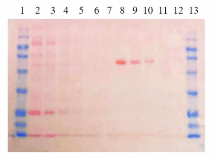
The
PVDF
membrane
stained
Ponceau
S
for
5
mins
and
washed
with
ddH2O
for
3
mins.
It
provides
visible
pink
bands.
Lane
1,13:
Prestained
MQ
Protein
Ladder
Lane
2
-
7:
2X
Dilutions
of
Unstained
Protein
Ladder
Lane
8
-
12:
2000,
1000,
500,
100,
50
ng
of
BSA
MQ
Ponceaus
S
Staining
Solution,
10X
concentrate
is
a
membrane
stain
for
evaluating
the
transfer
efficiency
of
a
western
blot.
This
stain
is
recommended
for
rapid
and
reversible
protein
staining
on
nitrocellulose
or
PVDF
membranes.
Ponceau
S
staining
is
reversible
and
can
be
removed
with
a
short
incubation
in
0.1%
NaOH.
Ponceau
S
solution
can
be
used
to
evaluate
for
total
protein
amount
or
transfer
efficiency
on
nitrocellulose
and
PVDF
membrane.
The
PVDF
membrane
stained
Ponceau
S
for
5
mins
and
washed
with
ddH2O
for
3
mins.
It
provides
visible
pink
bands.
Lane 1,
13:
Prestained
MQ
Protein
Ladder
Lane
2
-
7:
2X
Dilutions
of
Unstained
Protein
Ladder
Lane
8
-
12:
2000,
1000,
500,
100,
50
ng
of
BSA
MQ
Ponceaus
S
Staining
Solution
is
a
ready
-
to
-
use
membrane
stain
for
evaluating
the
transfer
efficiency
of
a
western
blot.
This
stain
is
recommended
for
rapid
and
reversible
protein
staining
on
nitrocellulose
or
PVDF
membranes.
Ponceau
S
staining
is
reversible
and
can
be
removed
with
a
short
incubation
in
0.1%
NaOH
.
Ponceau
S
solution
can
be
used
to
evaluate
for
total
protein
amount
or
transfer
efficiency
on
nitrocellulose
and
PVDF
membrane.
The
PVDF
membrane
stained
Ponceau
S
for
5
mins
and
washed
with
ddH2O
for
3
mins.
It
provides
visible
pink
bands.
Lane
1,13:
Prestained
MQ
Protein
Ladder
Lane
2
-
7:
2X
Dilutions
of
Unstained
Protein
Ladder
Lane
8
-
12:
2000,
1000,
500,
100,
50
ng
of
BSA
MQ
Ponceaus
S
Staining
Solution,
10X
concentrate
is
a
membrane
stain
for
evaluating
the
transfer
efficiency
of
a
western
blot.
This
stain
is
recommended
for
rapid
and
reversible
protein
staining
on
nitrocellulose
or
PVDF
membranes.
Ponceau
S
staining
is
reversible
and
can
be
removed
with
a
short
incubation
in
0.1%
NaOH.
Ponceau
S
solution
can
be
used
to
evaluate
for
total
protein
amount
or
transfer
efficiency
on
nitrocellulose
and
PVDF
membrane.
The
PVDF
membrane
stained
Ponceau
S
for
5
mins
and
washed
with
ddH2O
for
3
mins.
It
provides
visible
pink
bands.
Lane 1,
13:
Prestained
MQ
Protein
Ladder
Lane
2
-
7:
2X
Dilutions
of
Unstained
Protein
Ladder
Lane
8
-
12:
2000,
1000,
500,
100,
50
ng
of
BSA
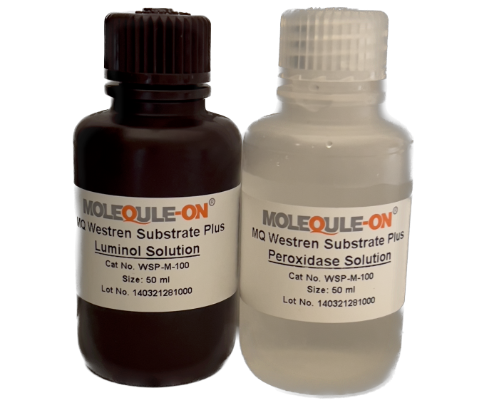
The
MQ
Western
Substrate
Plus
works
as
a
luminol
-
based
enhanced
chemiluminescent
substrate,
is
sensitive
and
compatible
with
conducting
immunoblots
with
horseradish
peroxidase
(HRP)
–
conjugated
secondary
antibodies.
The
low
picogram
or
high
femtogram
detection
of
antigen
is
enabled
by
MQ
Western
Substrate
Plus
shows
excellent
sensitivity
and
long
signal
duration.
Further,
its
long
chemiluminescent
signal
duration
makes
both
digital
and
film
-
based
imaging
possible
without
any
loss
of
the
signal.
Appropriate
primary
and
secondary
antibody
dilutions
are
suggested
for
attaining
optimal
signal
intensity
and
duration.
Features
1. No
optimization
required.
Switching
to
the
MQ
Western
Substrate
Plus
from
other
brands,
such
as
Pierce
ECL
and
GE
Healthcare,
does
not
require
optimization
or
protocol
changes.
2. High
degree
of
sensitivity
and
enhanced
chemiluminescence
duration.
MQ
Western
Substrate
Plus
enables
an
accurate
low
picogram
or
high
femtogram
detection
of
protein
on
the
same
immunoblot
after
a
single
exposure.
3. Optimized
for
use
with
PVDF
and
nitrocellulose
membranes.
4. Compatible
with
Western
Blotting
Markers.
5. Optimized
for
film
-
and
CCD
-
based
imaging.
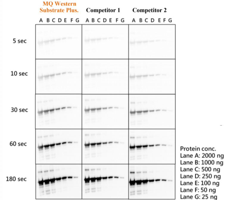
Low
picogram
to
high
femtogram
detection
MQ
Western
Substrate
Plus
enables
an
accurate
low
picogram
to
high
femtogram
detection
of
protein
on
the
same
immunoblot
after
a
single
exposure.
Membranes
were
probed
with
GFP
tag
Rabbit
Poly
Ab
diluted
at
1:10,000
of
and
then
with
Goat
Anti
-
rabbit
IgG/HRP
secondary
antibody
(1:10,000)
after
serial
dilution
EGFP
(Enhanced
Green
Fluorescent
Protein)
were
prepared
and
applied
in
electrophoresis
and
protein
transfer.
Identical
blots
were
incubated
with
the
Western
substrate.
The
blots
were
simultaneously
exposed
for
5
seconds,
10
seconds,
30
seconds,
60
seconds,
and
180
seconds
using
imaging
system.
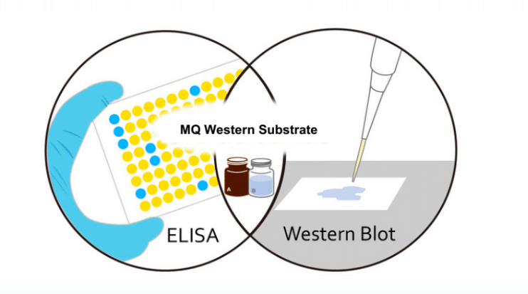
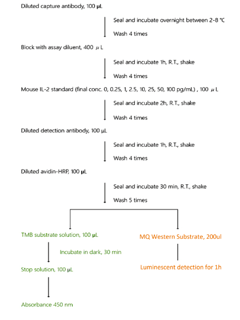
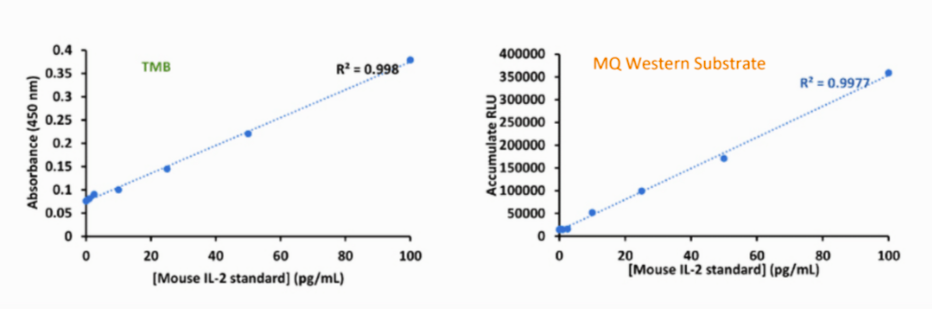
MQ Western Substrate can also be applied as ELISA substrates.
The
MQ
Western
Substrate
Ultra
works
as
a
luminol
-
based
enhanced
chemiluminescent
substrate,
is
sensitive
and
compatible
with
conducting
immunoblots
with
horseradish
peroxidase
(HRP)
–
conjugated
secondary
antibodies.
The
low
picogram
to
mid
femtogram
detection
of
antigen
is
enabled
by
MQ
Western
Substrate
Ultra
shows
excellent
sensitivity
and
long
signal
duration.
Further,
its
long
chemiluminescent
signal
duration
makes
both
digital
and
film
-
based
imaging
possible
without
any
loss
of
the
signal.
Appropriate
primary
and
secondary
antibody
dilutions
are
suggested
for
attaining
optimal
signal
intensity
and
duration.
Features
1. No
optimization
required.
Switching
to
the
MQ
Western
Substrate
Ultra
from
other
brands,
such
as
Pierce
ECL
and
GE
Healthcare,
does
not
require
optimization
or
protocol
changes.
2. High
degree
of
sensitivity
and
enhanced
chemiluminescence
duration.
MQ
Western
Substrate
Ultra
enables
an
accurate
low
picogram
or
high
femtogram
detection
of
protein
on
the
same
immunoblot
after
a
single
exposure.
3. Optimized
for
use
with
PVDF
and
nitrocellulose
membranes.
4. Compatible
with
Western
Blotting
Markers.
5. Optimized
for
film
-
and
CCD
-
based
imaging.
Low
picogram
to
mid
femtogram
detection
MQ
Western
Substrate
Ultra
enables
an
accurate
low
picogram
to
mid
femtogram
detection
of
protein
on
the
same
immunoblot
after
a
single
exposure.
Membranes
were
probed
with
GFP
tag
Rabbit
PolyAb
diluted
at
1:10,000
of
and
then
with
Goat
Anti
-
rabbit
IgG/HRP
secondary
antibody
(1:10,000)
after
serial
dilution
EGFP
(Enhanced
Green
Fluorescent
Protein)
were
prepared
and
applied
in
electrophoresis
and
protein
transfer.
Identical
blots
were
incubated
with
the
Western
substrate.
The
blots
were
simultaneously
exposed
for
5
seconds,
10
seconds,
30
seconds,
60
seconds,
and
180
seconds
using
imaging
system.
MQ Western Substrate can also be applied as ELISA substrates.
The
MQ
Western
Substrate
Plus
works
as
a
luminol
-
based
enhanced
chemiluminescent
substrate,
is
sensitive
and
compatible
with
conducting
immunoblots
with
horseradish
peroxidase
(HRP)
–
conjugated
secondary
antibodies.
The
low
picogram
or
high
femtogram
detection
of
antigen
is
enabled
by
MQ
Western
Substrate
Plus
shows
excellent
sensitivity
and
long
signal
duration.
Further,
its
long
chemiluminescent
signal
duration
makes
both
digital
and
film
-
based
imaging
possible
without
any
loss
of
the
signal.
Appropriate
primary
and
secondary
antibody
dilutions
are
suggested
for
attaining
optimal
signal
intensity
and
duration.
Features
1. No
optimization
required.
Switching
to
the
MQ
Western
Substrate
Plus
from
other
brands,
such
as
Pierce
ECL
and
GE
Healthcare,
does
not
require
optimization
or
protocol
changes.
2. High
degree
of
sensitivity
and
enhanced
chemiluminescence
duration.
MQ
Western
Substrate
Plus
enables
an
accurate
low
picogram
or
high
femtogram
detection
of
protein
on
the
same
immunoblot
after
a
single
exposure.
3. Optimized
for
use
with
PVDF
and
nitrocellulose
membranes.
4. Compatible
with
Western
Blotting
Markers.
5. Optimized
for
film
-
and
CCD
-
based
imaging.
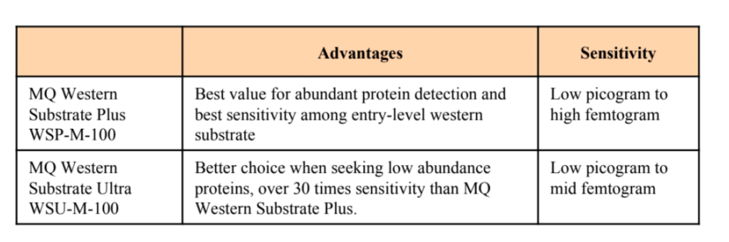
Low
picogram
to
high
femtogram
detection
MQ
Western
Substrate
Plus
enables
an
accurate
low
picogram
to
high
femtogram
detection
of
protein
on
the
same
immunoblot
after
a
single
exposure.
Membranes
were
probed
with
GFP
tag
Rabbit
Poly
Ab
diluted
at
1:10,000
of
and
then
with
Goat
Anti
-
rabbit
IgG/HRP
secondary
antibody
(1:10,000)
after
serial
dilution
EGFP
(Enhanced
Green
Fluorescent
Protein)
were
prepared
and
applied
in
electrophoresis
and
protein
transfer.
Identical
blots
were
incubated
with
the
Western
substrate.
The
blots
were
simultaneously
exposed
for
5
seconds,
10
seconds,
30
seconds,
60
seconds,
and
180
seconds
using
imaging
system.
MQ Western Substrate can also be applied as ELISA substrates.
The
MQ
Western
Substrate
Ultra
works
as
a
luminol
-
based
enhanced
chemiluminescent
substrate,
is
sensitive
and
compatible
with
conducting
immunoblots
with
horseradish
peroxidase
(HRP)
–
conjugated
secondary
antibodies.
The
low
picogram
to
mid
femtogram
detection
of
antigen
is
enabled
by
MQ
Western
Substrate
Ultra
shows
excellent
sensitivity
and
long
signal
duration.
Further,
its
long
chemiluminescent
signal
duration
makes
both
digital
and
film
-
based
imaging
possible
without
any
loss
of
the
signal.
Appropriate
primary
and
secondary
antibody
dilutions
are
suggested
for
attaining
optimal
signal
intensity
and
duration.
Features
1. No
optimization
required.
Switching
to
the
MQ
Western
Substrate
Ultra
from
other
brands,
such
as
Pierce
ECL
and
GE
Healthcare,
does
not
require
optimization
or
protocol
changes.
2. High
degree
of
sensitivity
and
enhanced
chemiluminescence
duration.
MQ
Western
Substrate
Ultra
enables
an
accurate
low
picogram
or
high
femtogram
detection
of
protein
on
the
same
immunoblot
after
a
single
exposure.
3. Optimized
for
use
with
PVDF
and
nitrocellulose
membranes.
4. Compatible
with
Western
Blotting
Markers.
5. Optimized
for
film
-
and
CCD
-
based
imaging.
Low
picogram
to
mid
femtogram
detection
MQ
Western
Substrate
Ultra
enables
an
accurate
low
picogram
to
mid
femtogram
detection
of
protein
on
the
same
immunoblot
after
a
single
exposure.
Membranes
were
probed
with
GFP
tag
Rabbit
PolyAb
diluted
at
1:10,000
of
and
then
with
Goat
Anti
-
rabbit
IgG/HRP
secondary
antibody
(1:10,000)
after
serial
dilution
EGFP
(Enhanced
Green
Fluorescent
Protein)
were
prepared
and
applied
in
electrophoresis
and
protein
transfer.
Identical
blots
were
incubated
with
the
Western
substrate.
The
blots
were
simultaneously
exposed
for
5
seconds,
10
seconds,
30
seconds,
60
seconds,
and
180
seconds
using
imaging
system.
MQ Western Substrate can also be applied as ELISA substrates.
Phosphate buffered saline (PBS) from MOLEQULE
-
ON
available
as a 1X
Working Solution.
It is
filtered
through 0.2 μm filter membrane.
The
buffer is isotonic and non
-
toxic to most cells
therefore,
it is used in many biological research procedures.
PBS Buffer commonly used as substance dilution,
cell wash solution and
rinsing of cell container.
Working Solution
In 1X solution, the concentration of phosphate buffer is 0.01M while of sodium chloride is 0.154M.
The solution pH will be ~7.2
-
7.4.
Phosphate buffered saline (PBS) from MOLEQULE
-
ON
available
as a 10X concentrate.
It is
filtered
through 0.2 μm filter membrane.
The
buffer is isotonic and non
-
toxic to most cells
therefore,
it is used in many biological research procedures.
PBS Buffer commonly used as
substance dilution, cell wash solution and
rinsing of cell container.
Working Solution
To make
one liter
of
1X working concentration
of PBS,
mix 100ml of 10X Phosphate B
uffered
Saline in 900ml of sterile distilled water. In 1X solution, the concentration of phosphate buffer is
0.01M while of sodium chloride is 0.154M. The solution pH will be ~7.2
- 7.4.
Phosphate buffered saline (PBS) from MOLEQULE
-
ON
available
as a 1X
Working Solution.
It is
filtered
through 0.2 μm filter membrane.
The
buffer is isotonic and non
-
toxic to most cells
therefore,
it is used in many biological research procedures.
PBS Buffer commonly used as substance dilution,
cell wash solution and
rinsing of cell container.
Working Solution
In 1X solution, the concentration of phosphate buffer is 0.01M while of sodium chloride is 0.154M.
The solution pH will be ~7.2
-
7.4.
Phosphate buffered saline (PBS) from MOLEQULE
-
ON
available
as a 10X concentrate.
It is
filtered
through 0.2 μm filter membrane.
The
buffer is isotonic and non
-
toxic to most cells
therefore,
it is used in many biological research procedures.
PBS Buffer commonly used as
substance dilution, cell wash solution and
rinsing of cell container.
Working Solution
To make
one liter
of
1X working concentration
of PBS,
mix 100ml of 10X Phosphate B
uffered
Saline in 900ml of sterile distilled water. In 1X solution, the concentration of phosphate buffer is
0.01M while of sodium chloride is 0.154M. The solution pH will be ~7.2
- 7.4.
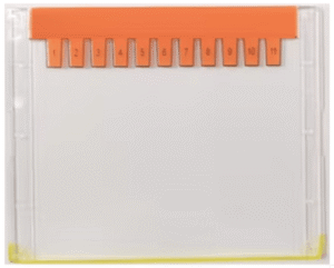
The MQ Easy Protein Pre-cast Gel is a convenient and quick-to-use polyacrylamide gel. The pre-cast gel plate has a unique design with a special surface treatment that improves protein band resolution for a more even distribution of bands. The gel is based on the Tris system and works well with Tris-glycine SDS-PAGE standard electrophoresis buffer. Additionally, the product includes two bags of Tris-glycine SDS-PAGE high-resolution rapid electrophoresis buffer powder. This high-resolution rapid electrophoresis buffer has a strong buffering capacity and provides higher resolution, resulting in sharper and clearer protein bands. It does not contain SDS and can be used for native electrophoresis with appropriate electrophoresis buffers and corresponding reagents.
Specification
1. No.
of
wells:
11 or 15
wells
2. Percentage: 4-12%
3. Separation range: 10 kDa to 300 kDa
4. Gel plate dimension: 8.2 cm x 10 cm
5. Maximum loading volume of gel well: 40 μl
6. Buffer: Tris-Glycine
Compatibility
1. Bio-Rad Mini-PROTEAN® II & 3
2. Bio-Rad Mini-PROTEAN® Tetra System
3. MQ 2-Gels Vertical Gel Electrophoresis Unit
4. MQ 4-Gels Vertical Gel Electrophoresis Unit
MQ
Western
One
Step
Blocker
is
a
blocking
solution
for
Western
blot
analysis.
This
Blocker
buffer
not
only
provides
blocking
and
primary
and
secondary
antibody
hybridization
in
one
step
but
also
enhances
the
signal
developed
with
HRP
(horseradish
peroxidase)
or
AP
(alkaline
phosphatase)
substrates.
It,
therefore,
serves
as
both
blocker
and
enhancer
in
Western
analysis.
With
the
three
-
in
-
one
step
procedure,
MQ
Western
One
Step
Blocker
is
a
time
and
labor
economic
solution
for
the
time
consuming
and
laborious
Western
procedure.
Features
3
Steps
in
One:
Block
the
membrane
and
dilute
primary
and
secondary
antibody
in
one
step.
Enhance
antibody
signal:
It
shows
a
two-to
five-fold
increase
in
signal
intensity
for
most
protein
targets,
enabling
much
less
protein
to
be
detected
with
the
same
substrate
and
method.
Enhance
time-saving:
It
saves
at
least
2
hours
in
the
antibody
detection
process
during
the
Western
Blot,
with
only
one
hour
needed.
Universal
antibody
diluent:
Ready-to-use
dilution
buffer
for
most
of
primary
and
secondary
antibody.
No
blocking
step
needed:
Just
immerse
the
membrane
in
the
MQ
Western
One
Step
Blocker
solution
with
your
antibodies.
Effective
with
any
ECL:
After
the
antibody
detection
process,
the
signal
can
be
developed
with
both
HRP
(horseradish
peroxidase)
and
AP
(alkaline
phosphatase)
substrates.
Less
hands-on
steps:
No
3
wash
steps
are
required,
meaning
no
need
to
transfer
the
membrane
in
&
out
of
the
container.
Compatible
with
PVDF
&
NC
membrane:
Regardless
of
the
pore
size,
the
MQ
Blocker
minimizes
the
background
from
non
-
specific
protein
binding
by
antibodies.
Improve
protein
detection:
Improve
the
binding
process
of
target
proteins,
so
that
specific
antibodies
can
bind
more
effectively.
Protein
free:
Reduces
overall
background
and
minimizes
non-specific
signals
often
seen
with
ECL
detection.
1°Ab
(ab
8227,
abcam
):
Rabbit
anti
beta
actin;
1:5,000
2°Ab
(ab
205718,
abcam
):
Goat
anti
Rabbit
HRP;
1:5,000
Nitrocellulo
se and PVDF membranes probed by
Western blotting procedures and detected by
chemiluminescent
or other non
-
precipitating substrates can be stripped and re
-
probed using
MQ
Western
Stripping Buffer. Western blotting is widely used to detect and compare proteins in complex
mixtures, and
chemiluminescence has largely replaced chromogenic substrates as
the most
convenient and sensitive
method of detection. One advantage of chemiluminescence is the ability to
strip and re
-
probe the protein
mixture on the membrane. Traditional stripping methods use conditions
that are effective for only low
-
affinity
antibody
-
antigen interactions or are so harsh that they tend to
adversely alter the antigen for
subsequent immunoprobing. MQ
Western Stripping Buffer
is a robust but gentle formulation for stripping
primary and secondary antibodies from blots to enable
several reprobings on the same membrane.
Tris Buffered Saline (TBS) is a
buffering
solution
that stabilizes
pH. The solution is commonly used
for
western blot and ELISA procedures. It is used as a convenient prepared buffer for use in various
applications.
Working Solution
To make one liter of 1X working concentration, take 50ml of 20X
TBS
Buffer and mix with 950ml
of sterile distilled water. 1X
Buffer contains 0.14M Sodium Chloride, 3mM Potassium Chloride and
25mM Tris Base.
Lorem ipsum dolor sit amet, consectetur adipisicing elit. Optio, neque qui velit. Magni dolorum quidem ipsam eligendi, totam, facilis laudantium cum accusamus ullam voluptatibus commodi numquam, error, est. Ea, consequatur.
Ponceau S Protein Staining Solution
Lorem ipsum dolor sit amet, consectetur adipisicing elit. Optio, neque qui velit. Magni dolorum quidem ipsam eligendi, totam, facilis laudantium cum accusamus ullam voluptatibus commodi numquam, error, est. Ea, consequatur.
Western chemiluminescence Substrate
Lorem ipsum dolor sit amet, consectetur adipisicing elit. Optio, neque qui velit. Magni dolorum quidem ipsam eligendi, totam, facilis laudantium cum accusamus ullam voluptatibus commodi numquam, error, est. Ea, consequatur.
PBS Buffer
Lorem ipsum dolor sit amet, consectetur adipisicing elit. Optio, neque qui velit. Magni dolorum quidem ipsam eligendi, totam, facilis laudantium cum accusamus ullam voluptatibus commodi numquam, error, est. Ea, consequatur.
Easy Protein Gels
Lorem ipsum dolor sit amet, consectetur adipisicing elit. Optio, neque qui velit. Magni dolorum quidem ipsam eligendi, totam, facilis laudantium cum accusamus ullam voluptatibus commodi numquam, error, est. Ea, consequatur.
MQ Western One Step Blocker
Lorem ipsum dolor sit amet, consectetur adipisicing elit. Optio, neque qui velit. Magni dolorum quidem ipsam eligendi, totam, facilis laudantium cum accusamus ullam voluptatibus commodi numquam, error, est. Ea, consequatur.
MQ Western Stripping Buffer
Lorem ipsum dolor sit amet, consectetur adipisicing elit. Optio, neque qui velit. Magni dolorum quidem ipsam eligendi, totam, facilis laudantium cum accusamus ullam voluptatibus commodi numquam, error, est. Ea, consequatur.
Tris Buffered Saline Buffer, TBS, (20X)
Lorem ipsum dolor sit amet, consectetur adipisicing elit. Optio, neque qui velit. Magni dolorum quidem ipsam eligendi, totam, facilis laudantium cum accusamus ullam voluptatibus commodi numquam, error, est. Ea, consequatur.